
Cranial Nerve Examination: A Comprehensive Guide
This guide details systematic assessment of each cranial nerve, identifying abnormalities linked to neurological pathologies, offering essential knowledge for comprehensive neurological evaluations.
Cranial nerve examination is a fundamental component of the neurological assessment, providing crucial insights into the function of the peripheral nervous system. This systematic evaluation assesses each of the twelve cranial nerves, revealing potential locations of lesions impacting neurological pathways. A thorough examination involves testing specific functions associated with each nerve, like smell, vision, and facial movement.
Understanding these nerves and their functions is vital for diagnosing a wide range of neurological conditions, from stroke and tumors to multiple sclerosis and Bell’s palsy. Accurate interpretation of findings requires a solid grasp of neuroanatomy and clinical correlation.
Importance of Cranial Nerve Assessment
Cranial nerve assessment is paramount in localizing neurological deficits, aiding in differential diagnosis and guiding further investigations. Abnormalities detected can pinpoint lesions within the brainstem, cerebellum, or along the course of individual nerves. Early identification of cranial nerve dysfunction is crucial for timely intervention and improved patient outcomes.
This assessment helps differentiate between central and peripheral nervous system disorders, informing treatment strategies. A detailed examination provides baseline data for monitoring disease progression or response to therapy, ultimately enhancing patient care and neurological understanding.
Tools and Equipment for Examination
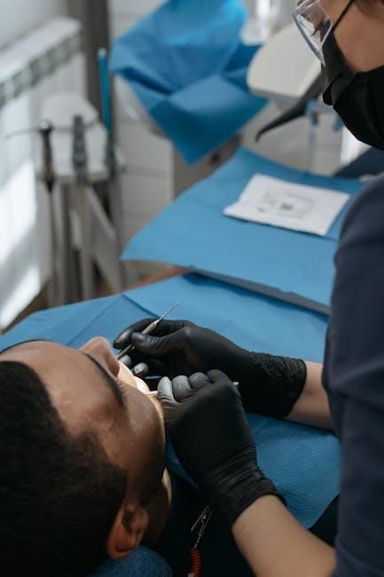
A comprehensive cranial nerve examination requires minimal specialized equipment. Essential tools include a penlight for pupillary responses and facial sensation testing, and an ophthalmoscope for fundoscopic examination of the optic disc. Additionally, tuning forks (Weber and Rinne) are needed for auditory assessment.
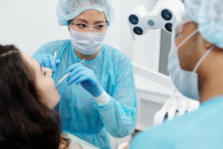
Cotton-tipped applicators are utilized for corneal reflex testing, while materials for olfactory stimulation (coffee, vanilla) are beneficial. A tongue depressor aids in assessing gag reflex and tongue movement. Careful observation and patient history remain the most vital components.

Detailed Examination of Individual Cranial Nerves
This section provides a systematic approach to evaluating each of the twelve cranial nerves, detailing specific tests and expected findings for accurate neurological assessment.
I. Olfactory Nerve (CN I) – Smell
Assessing the olfactory nerve involves testing a patient’s ability to identify common odors, utilizing non-irritating substances in each nostril independently. This evaluation helps pinpoint potential deficits in smell perception. Common disorders include anosmia (loss of smell), hyposmia (reduced smell), and parosmia (distorted smell).
These conditions can stem from nasal obstruction, neurological diseases, or head trauma. Careful documentation of patient responses and consideration of potential confounding factors are crucial for accurate diagnosis and management. A comprehensive olfactory assessment is a vital component of a thorough cranial nerve examination.
Testing Olfactory Function
Olfactory function testing requires presenting familiar, non-irritating odors – like coffee, vanilla, or lemon – to each nostril separately, while the patient keeps eyes closed. The examiner should observe the patient’s response, noting any difficulty identifying the scent or differences between nostrils.
It’s essential to avoid substances that stimulate the trigeminal nerve (like ammonia) as this can create a false positive. Documenting the intensity of the odor used and the patient’s accuracy is key for reliable results. This systematic approach ensures a precise evaluation of olfactory capabilities.
Common Olfactory Nerve Disorders
Anosmia, the complete loss of smell, is a frequent disorder, often stemming from nasal obstruction, upper respiratory infections, or head trauma. Hyposmia represents a reduced ability to smell, while parosmia involves distorted odor perception – familiar smells are recognized incorrectly.
Phantosmia, or olfactory hallucinations, presents as smelling odors that aren’t actually present. These disorders can significantly impact quality of life, affecting taste and potentially indicating underlying neurological conditions requiring further investigation and diagnosis.
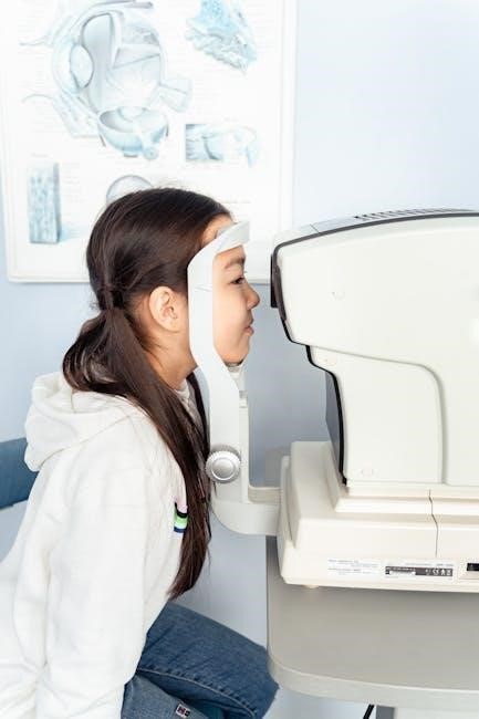
II. Optic Nerve (CN II) – Vision
The optic nerve transmits visual information from the retina to the brain, crucial for visual acuity, color perception, and pupillary responses. Assessing this nerve involves a series of tests designed to identify any disruptions in visual pathways.
Comprehensive evaluation includes visual acuity testing, visual field assessment to detect peripheral vision loss, and fundoscopic examination to inspect the optic disc for signs of damage or swelling, providing insights into overall neurological health.
Visual Acuity Testing
Visual acuity testing determines the sharpness of a patient’s vision, typically using a Snellen chart. Patients read lines of decreasingly smaller letters, indicating their ability to discern details at a distance.
Each eye is tested individually, both with and without corrective lenses, documenting the smallest line the patient can accurately read. Results are recorded as a fraction (e.g., 20/20), representing vision at 20 feet compared to standard vision.
Visual Field Assessment
Visual field assessment maps the extent of a patient’s peripheral vision, detecting deficits that may indicate optic nerve or brain damage. This is often performed using confrontation testing, where the examiner compares the patient’s visual field to their own.
More precise testing utilizes perimetry, presenting visual stimuli and recording the patient’s responses. Abnormalities, like scotomas (blind spots), can pinpoint the location of lesions affecting the visual pathway.
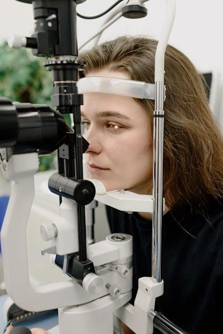
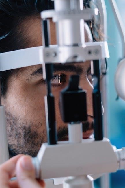
Fundoscopic Examination
Fundoscopic examination, utilizing an ophthalmoscope, allows direct visualization of the optic disc, retina, and blood vessels within the eye. This crucial step assesses the health of the optic nerve (CN II) and identifies potential pathologies.
Key findings include papilledema (swelling of the optic disc), optic atrophy, and retinal hemorrhages, all indicative of neurological conditions. Careful observation of the vascular structures can reveal signs of hypertension or diabetes.
III. Oculomotor Nerve (CN III) – Eye Movement
The oculomotor nerve controls most extraocular movements, specifically elevation, depression, and adduction of the eye. Assessment involves observing the patient’s ability to follow a moving target in all directions of gaze. Ptosis (drooping eyelid) and diplopia (double vision) are key indicators of CN III dysfunction.
Evaluation also includes assessing pupillary responses to light, as CN III innervates the pupillary constrictor muscle. A dilated, non-reactive pupil suggests potential compression of the nerve.
Pupillary Light Reflex
The pupillary light reflex tests afferent input via CN II (optic nerve) and efferent output through CN III (oculomotor nerve). Shining a light into one eye should cause constriction of both pupils – a consensual response.
Absence of direct or consensual response indicates a problem with either nerve. A sluggish response can suggest partial nerve damage or underlying systemic issues. Careful observation is crucial, noting pupil size and shape alongside the reflex response for accurate assessment.
Extraocular Muscle Function
Assessing extraocular muscle function evaluates CN III (oculomotor), IV (trochlear), and VI (abducens) nerves. The examiner observes eye movements in six cardinal directions of gaze, noting any weakness, nystagmus, or pain.
Diplopia (double vision) can indicate muscle weakness or nerve dysfunction. Smooth, coordinated movements are normal; limitations suggest a specific cranial nerve impairment. Documenting any deficits precisely is vital for accurate neurological localization and diagnosis.
IV. Trochlear Nerve (CN IV) – Eye Movement
The trochlear nerve innervates the superior oblique muscle, responsible for intorsion, depression, and external rotation of the eye. Assessing this nerve focuses on these specific movements. A lesion typically causes vertical diplopia that worsens when looking down and towards the opposite side of the lesion.
Testing involves observing eye movement in all directions of gaze, specifically noting any limitations in downward gaze. Careful observation is crucial for identifying subtle deficits indicative of trochlear nerve pathology.
Superior Oblique Muscle Assessment
Evaluating the superior oblique muscle is key to assessing trochlear nerve (CN IV) function. Patients are asked to look downwards and inwards; weakness presents as limited depression and intorsion of the eye. Observe for diplopia, particularly worsening with downward gaze.
The examiner should also assess for head tilt, a compensatory mechanism often adopted by patients with trochlear nerve palsy. This tilt helps minimize the double vision by aligning the visual axes. Careful observation is paramount.
V. Trigeminal Nerve (CN V) – Facial Sensation & Muscles
The trigeminal nerve governs facial sensation and motor function. Assessment begins with light touch testing across the three divisions: ophthalmic, maxillary, and mandibular. Note any asymmetry or numbness. Motor function is evaluated by palpating the masseter and temporalis muscles during clenching.
The corneal reflex tests afferent limb integrity; a diminished or absent reflex suggests nerve damage. Thorough evaluation identifies lesions impacting sensation or mastication, crucial for diagnosis.
Sensory Testing (Facial Regions)
Sensory evaluation of the trigeminal nerve divides into three regions: ophthalmic, maxillary, and mandibular. Use light touch with a cotton wisp, comparing bilaterally. Assess the forehead (ophthalmic), cheek (maxillary), and jaw (mandibular). Pinprick can assess pain sensation, though caution is advised.
Document any areas of diminished sensation, hyperesthesia, or allodynia. Precise localization helps pinpoint lesion sites. Patient reports of altered sensation are vital; carefully note their descriptions.
Motor Testing (Masseter & Temporalis)
Evaluate the masseter and temporalis muscles for strength, indicators of CN V motor function. Palpate the masseter as the patient clenches their teeth; note muscle bulk and tone. Assess temporalis strength by resisting upward and backward movement of the jaw.
Compare strength bilaterally, documenting any weakness or asymmetry. Reduced strength suggests CN V pathology. Observe for jaw deviation during clenching, another sign of weakness. Document findings clearly, noting any fasciculations or atrophy.
Corneal Reflex
Assess the corneal reflex to evaluate afferent (CN V) and efferent (CN VII) pathways. Gently touch the cornea with a wisp of cotton, observing for bilateral blink. A diminished or absent reflex indicates potential dysfunction. Ensure no prior corneal abrasion exists before testing.
Document the presence or absence of the reflex and any asymmetry. Absent reflex can signify CN V or VII damage. Note that the reflex tests both nerves, requiring careful interpretation. Always prioritize patient comfort and safety during this sensitive examination.
VI. Abducens Nerve (CN VI) – Eye Movement
The abducens nerve controls the lateral rectus muscle, responsible for abduction – moving the eye outward. Assess this function by asking the patient to follow a target horizontally, observing for full lateral movement in each eye. Diplopia (double vision) with lateral gaze suggests CN VI palsy.
Carefully note any limitations or weakness in abduction. Isolated CN VI palsy is common, often due to vascular or compressive issues. Document the degree of abduction deficit and any associated nystagmus. Thorough assessment is crucial for accurate neurological diagnosis.
Lateral Rectus Muscle Assessment
To assess the lateral rectus, instruct the patient to fixate on a distant target while you hold a penlight or finger. Ask them to follow the stimulus as you move it horizontally, observing each eye’s ability to abduct – move outwards. Weakness indicates potential CN VI dysfunction.
Note any diplopia (double vision) reported during abduction. Compare the range of motion between both eyes; asymmetry is significant. Document the degree of weakness and presence of nystagmus. Precise observation aids in pinpointing the neurological source of impairment.
VII. Facial Nerve (CN VII) – Facial Expression & Taste
Evaluating CN VII involves observing facial symmetry at rest and during voluntary movements. Ask the patient to smile, frown, raise eyebrows, and close eyes tightly. Note any asymmetry or weakness in these actions. Assess the nasolabial fold – flattening suggests facial nerve palsy.
Taste sensation testing focuses on the anterior two-thirds of the tongue using sweet, sour, or salty solutions. Document any taste discrimination deficits. A comprehensive assessment helps localize lesions affecting facial expression and gustatory function.
Facial Muscle Assessment (Smile, Frown, etc.)
Careful observation of facial movements is crucial for assessing CN VII function. Instruct the patient to perform specific actions: smile to evaluate the zygomaticus major, frown to test the corrugator supercilii, and tightly close their eyes, checking for complete closure. Observe for any asymmetry or weakness during these maneuvers.
Pay attention to the nasolabial fold; a diminished or flattened fold on one side can indicate facial nerve impairment. Document any involuntary movements or fasciculations. This detailed assessment helps pinpoint the location and extent of facial nerve dysfunction.
Taste Testing (Anterior 2/3 of Tongue)
Assessing taste on the anterior two-thirds of the tongue evaluates CN VII function. Use cotton swabs dipped in solutions representing sweet, sour, salty, and bitter tastes – ensuring solutions are not temperature-sensitive. Present each taste bilaterally, asking the patient to identify it with eyes closed.
Document the patient’s ability to correctly identify each taste. Diminished or absent taste sensation on one side suggests facial nerve damage. Control for nasal congestion, as smell influences taste perception. Accurate taste identification is vital for a complete neurological evaluation.
VIII. Vestibulocochlear Nerve (CN VIII) – Hearing & Balance
Evaluating CN VIII involves assessing both auditory and vestibular functions. Hearing is initially screened with whispered voice tests, followed by Weber and Rinne tests utilizing a tuning fork to differentiate conductive versus sensorineural hearing loss. Balance assessment includes the Romberg test, observing for loss of balance with eyes closed.
Further evaluation may include observing for nystagmus and performing Dix-Hallpike maneuver to assess for benign paroxysmal positional vertigo (BPPV). Document any deficits in hearing or balance, as these can indicate vestibulocochlear nerve dysfunction.
Hearing Assessment (Weber & Rinne Tests)
The Weber test assesses lateralization of sound. A vibrating tuning fork is placed on the midline of the head; normally, sound is perceived equally in both ears. Lateralization to one ear suggests conductive hearing loss on that side. The Rinne test compares air and bone conduction.
A vibrating tuning fork is placed on the mastoid bone, then near the ear canal. Normally, air conduction is greater than bone conduction (AC > BC). BC > AC indicates conductive hearing loss, while AC > BC suggests sensorineural loss.
Balance Testing (Romberg Test)
The Romberg test evaluates the contribution of proprioception and vestibular function to balance. The patient stands with feet together, arms at sides, and eyes open. Observe for swaying or loss of balance. Then, the patient closes their eyes.
Increased swaying or falling with eyes closed suggests a problem with proprioceptive or vestibular pathways. A positive Romberg sign indicates impaired balance control, potentially due to vestibular, proprioceptive, or cerebellar dysfunction. Careful observation is crucial for accurate assessment.

Advanced Considerations
Further evaluation may involve imaging or specialized tests to pinpoint the precise location and nature of cranial nerve dysfunction, aiding diagnosis.
IX. Glossopharyngeal Nerve (CN IX) – Swallowing & Taste
Assessing the glossopharyngeal nerve involves evaluating crucial functions like swallowing and taste perception. The gag reflex is a primary test, observing palatal elevation and pharyngeal constriction upon posterior tongue stimulation.
Taste testing focuses on the posterior third of the tongue, differentiating it from the facial nerve’s anterior two-thirds assessment. Dysfunction can manifest as difficulty swallowing (dysphagia) or altered taste sensation. Careful observation of these responses provides valuable diagnostic insight into potential neurological issues affecting this nerve.
Gag Reflex
The gag reflex is a critical component of the glossopharyngeal (CN IX) and vagus (CN X) nerve assessment. It tests the afferent limb of CN IX and the efferent limb of CN X.
Stimulation of the posterior pharyngeal wall, or slightly touching the back of the throat, should elicit a reflexive contraction of the palatoglossus and pharyngeal muscles, causing the palate to elevate and a gag response. Absence or asymmetry suggests neurological dysfunction, potentially indicating lesions along either nerve’s pathway, requiring further investigation.
Taste Testing (Posterior 1/3 of Tongue)
Assessing taste in the posterior third of the tongue evaluates the function of the glossopharyngeal nerve (CN IX). This is performed by applying small amounts of sugar or salt solutions to the back of the tongue using a cotton swab.
Patients should identify the taste; diminished or absent sensation on one side compared to the other indicates potential CN IX dysfunction. It’s crucial to ensure nasal passages are clear, as smell can influence taste perception, leading to inaccurate results during this neurological assessment.
X. Vagus Nerve (CN X) – Swallowing, Speech & Autonomic Function
The vagus nerve profoundly impacts swallowing, speech, and crucial autonomic functions. Examination involves assessing palate elevation during phonation (“ah” sound) and observing for symmetry. A deviated uvula suggests weakness.
Gag reflex testing, stimulating the posterior pharynx, evaluates afferent and efferent vagal pathways. Dysphagia or hoarseness can indicate vagal nerve impairment. Assessing heart rate variability also provides insight into vagal tone and autonomic control, completing the comprehensive evaluation.
Assessment of Palate and Pharynx
Palate and pharyngeal assessment is critical for evaluating Vagus nerve (CN X) function. Observe for symmetrical elevation of the soft palate during phonation, specifically when the patient says “Ah.” Asymmetry suggests unilateral weakness.
Note the presence of a deviated uvula, which also indicates impaired palatal muscle function. Carefully examine the pharynx for any signs of asymmetry or reduced movement during swallowing, potentially signaling neurological deficits affecting speech and deglutition.
XI. Accessory Nerve (CN XI) – Shoulder Shrugging & Head Turning
The Accessory Nerve (CN XI) controls the trapezius and sternocleidomastoid muscles. Assess shoulder shrug against resistance; weakness indicates accessory nerve dysfunction. Observe head turning against resistance, evaluating sternocleidomastoid strength bilaterally.
Palpate the muscles for atrophy or fasciculations. Document any asymmetry in muscle bulk or strength. Thorough testing helps pinpoint lesions affecting this nerve, impacting neck and shoulder movement, crucial for a complete neurological assessment.
Trapezius and Sternocleidomastoid Muscle Testing
Evaluate trapezius strength by asking the patient to shrug their shoulders against resistance. Note any weakness or asymmetry. Assess sternocleidomastoid function by having the patient turn their head to each side against opposing force.
Observe for full range of motion and equal resistance. Palpate both muscles for tone, bulk, and any signs of fasciculations. Document findings meticulously, as deficits indicate potential CN XI impairment, impacting neck and shoulder functionality.
XII. Hypoglossal Nerve (CN XII) – Tongue Movement
Observe the tongue at rest for any fasciculations or asymmetry. Ask the patient to protrude their tongue, noting the midline position and any deviation.
Then, request lateral movements, assessing the range of motion on both sides. Weakness or deviation suggests CN XII dysfunction. Document observations carefully, as this nerve controls tongue musculature crucial for speech and swallowing. A thorough assessment aids in diagnosing neurological conditions affecting motor control.
Tongue Protrusion and Lateral Movement
Instruct the patient to protrude their tongue, observing for midline deviation. Any asymmetry during protrusion indicates potential hypoglossal nerve weakness. Subsequently, ask the patient to move the tongue laterally from side to side against resistance.
Assess the range of motion and strength of lateral movements. Difficulty or weakness suggests impairment of CN XII function. Document any observed fasciculations or atrophy. This detailed evaluation helps pinpoint the location and severity of nerve damage.